Dallas
214-456-2240
Fax: 214-456-8881
Plano
469-497-2501
Fax: 469-497-2507
Hemangiomas are benign tumors made up of dilated blood vessels that typically disappear over time.
214-456-2240
Fax: 214-456-8881
469-497-2501
Fax: 469-497-2507
Hemangiomas, also known as Infantile Hemangiomas, are the most common tumor of infancy, affecting approximately 5% of all infants and increases to 10 – 12% by one year of age. These tumors are usually found in the head and neck area, and occur most frequently in female premature infants with low birth weight.
There are many outdated terms for hemangiomas that are still commonly used. In the past, different terms were used to describe hemangiomas based upon their appearance which really depends on what depth they are growing in relation to the skin.
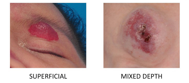
Superficial Hemangiomas are located in the skin, previously known as Strawberry Hemangiomas.
Deep Hemangiomas are located directly under the skin, previously known as Cavernous Hemangiomas.
Mixed Depth Hemangiomas are located in and under the skin with mixed level involvement, previously known as Capillary Cavernous Hemangiomas.

It is important to remember that the old terms all refer to the same tumor (hemangiomas) that behave the same and are benign. The only difference is that they occur at different depths in the skin and this gives them a different appearance.
Nasal hemangiomas are particularly worrying for parents, as the bright red area is obvious on a clearly visible part of the face, and covering or hiding it is difficult. In addition, large hemangiomas can block the air passages within the nose and can distort the underlying nasal structure, which can lead to ongoing deformity even after the hemangioma has involuted. Our approach to treating nasal hemangiomas is to use the appropriate combination of all hemangioma treatment types considering the age of the patient, and the site, size and effect of the hemangioma on the rest of the nose. We offer medical therapy such as Propranolol, different types of LASER, and surgery both to remove the swelling and to reconstruct the nose to give the overall best long-term result for the child.
Significant deformity of the specialized tissues such as cartilage in the nose and ear, the red portion of the lips or eyelid structures that are very difficult to reconstruct mandate a more aggressive treatment strategy than hemangiomas occurring in the trunk or extremities. Often times using oral medications is needed to attempt to limit growth and/or shrink hemangiomas that are close to specialized structures or area that present a functional concern. About 20% of infants will present with more than one hemangioma. If there are less than 5 tumors there are no special considerations. In rare circumstances, an infant may be found to have many hemangiomas (more than 5), which raises concern that hemangiomas may be found in the internal organs as well. The liver is the most common place that hemangiomas are found. It should be noted that incidental finding of hemangiomas in the liver is common. The liver hemangiomas to be concerned about are the ones that are very large because they can place extra stress on the heart and other problems that should be treated aggressively. While these are instances are very rare, patients with more than 5 lesions should have a liver ultrasound to ensure a large hemangioma of the liver is not present.
Occasionally the skin involved in the tumor will break down and open. This occurs because the damage to the skin by the tumor weakens it. This skin is further weakened by having a poor blood supply which slows healing. This makes the skin in the area of hemangiomas very delicate and even minor trauma or exposure to damp conditions to cause the skin to open. Ulceration occurs most often in the diaper area, but may occur anywhere. Preventive use of barrier cream in the diaper area to shield the hemangioma from moisture, and use of moisturizer in all other areas helps to maintain supple and resilient skin and reduces the chance of ulceration. When ulceration does occur, the wound care plan is prescribed according to the location and depth of the ulceration and this should be prescribed by an experienced plastic surgeon.
PHACE is an acronym with each letter standing for a constellation of findings that are found in about 2% of patients with hemangiomas. “P” stands for posterior brain malformation. “H” stands for hemangioma. “A” stands for abnormal arteries in the brain. “C” stands for cardiac (heart) abnormalities and coarctation (narrowing) of the aorta. “E” stands for eye and endocrine (hormone) abnormalities. Nine out of Ten patients with PHACE are female. The greatest concern for infants suspected of having PHACE is related to the risk for stroke (8%) related to the abnormal blood vessels (70%) and about 40% have some kind of malformation of the brain. MRIs are obtained in patients suspected of having PHACE syndrome to detect these common and serious anomalies.
Congenital (present at birth) hemangiomas are rare. They are present and are already enlarged at birth and usually have a different appearance from infantile hemangiomas. They have a pale base with the purple spots of color (stippling), but occasionally have a similar appearance to infantile hemangiomas. The rapidly involuting type (RICH) shrinks and fades (involutes) shortly after birth. These are not just “early infantile hemangiomas.” These tumors are made up of different cells than infantile hemangiomas but they are benign and follow a similar time course and treatment strategy.

These hemangiomas are also present at birth but unlike RICH these do not involute, nor do they grow. NICHs stay similar in size and appearance over time. These tumors (see picture above) have a different appearance from infantile hemangiomas. Most commonly, these have a pale base with the purple spots of color (stippling).

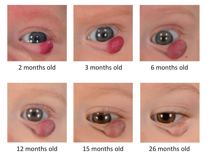
This involution phase is a period when the tumor cells are replaced with fatty and scar tissue. As seen above, the onset of involution is accompanied by a grey color change to the skin of the hemangioma and a significant softening of the tumor – becoming more spongy. This period continues over the course of years and is usually complete by age 7 but may last until ten years of age. After involution, half of children will not have any noticeable abnormality in the area where the hemangioma was located. In the other half of patients, there can be crepe like (paper thin) skin, excess skin that hangs, residual extra bulk in and under the skin or deformity of structures under the hemangioma (such as in the nose or ear where cartilages may be damaged by the hemangioma).
Hemangiomas are so common and have such a characteristic growth pattern that the history of how the tumor appeared and the clinical exam are usually all that experienced physicians need to make the diagnosis. Hemangiomas are typically diagnosed by appearance, but if the diagnosis is not clear a biopsy can be used to confirm the diagnosis, however this is rare.
While all hemangiomas are benign, there are special cases when the location of the hemangioma and associated patterns that accompany them need to be recognized. The first thing to understand is that despite being benign, non invasive tumors, the bulk of hemangiomas can deform delicate specialized structures and create difficult reconstructive challenges. This is most common in the head and neck. Hemangiomas in the eyelids, nose, lip and ear can irreversibly damage underlying cartilage, pigmentation or skin quality that will result in a deformity of these structures. We must also consider functional problems in the eyelids that can cause problems with vision. If large, the weight of the hemangioma can cause the upper eyelid to close and block the pupil. If the hemangioma is very large it can block the vision directly. In infants, if the brain is not receiving a visual picture from an eye the brain will begin to ignore the information coming from that eye (deprivation amblyopia). This makes it important to treat hemangiomas in this area with medication or surgery early. If very large, hemangiomas can compress the eye that can cause the eye to become misshapen (astigmatism). Another important location is in the beard distribution (along the jawline, chin and neck) as these hemangiomas can compress the windpipe (trachea).
It is not well understood why hemangiomas occur. There are no known associations between maternal diet, environment or behaviors.
Observation (no treatment) has been, and will continue to be, the treatment of choice in most patients with hemangiomas. This is because half of hemangiomas leave no scarring or evidence that they were there, and often the visible changes after involution are subtle. This is particularly true in less cosmetically sensitive areas such as the trunk and extremities.
A minimally invasive treatment that reduces blood flow, with potential surgical removal of the hemangioma if necessary.
For many years, intralesional injection of steroids or oral steroid treatment were the main medications used for treatment of hemangiomas. Intralesional steroids can slow the growth of hemangiomas and potentially shrink them, but the effects of these injections last for only 4-6 weeks. This means that injections would be needed every 4-6 weeks. There are also side effects from the steroids including thinning of the skin and loss of pigmentation in surrounding tissues that limits the use of this treatment. If the hemangioma is close to an eye injection of steroid is associated with a very small risk of blindness. Oral steroids are effective and generally considered safe. However, the common side effects accompanying steroid treatment include puffing up of the cheeks giving a bloated appearance (Cushingoid features), irritability and stomach upset. Other side effects include decreased height (although this is temporary) and elevation of the blood pressure. This historically led to low rates of prescribing steroids except for extreme cases.
Recently propranolol has been introduced for use in treating hemangiomas. Propranolol is a beta blocker that was originally created for treating conditions requiring blood pressure and heart rate control. The incidental benefit of causing hemangiomas to decrease in size was discovered in infants treated with propranolol who happened to have hemangiomas as well. Since that time, several case series have been published illustrating a good safety profile and efficacy of this drug. The side effects include potential for low blood pressure, low heart rate and decreased blood sugar levels. From our experience, these side effects occur rarely and are monitored for to ensure that they do not occur and are detected early if they do occur. The medication is started at half of the therapeutic dose to allow a gentle transition and avoid problems with side effects. The blood pressure and heart rate and general wellness of the patient are monitored. If tolerated, the full dose is then initiated and the same monitoring process is performed.
In general, the vast majority of patients tolerate the medication. We continue to monitor blood pressure and heart rate and general wellness monthly. The medication is continued until 12-18 months of age based upon the location and depth of the hemangioma. Occasionally, deep hemangiomas may begin growing again, and if this occurs the medication is restarted. In our experience, the rate of side effects is low allowing us to prescribe propranolol for hemangiomas that we would have avoided prescribing steroids for in the past.
The role of using Pulsed dye laser or similar lasers (532 nm or 595 nm lasers) is essentially reserved for the involution phase to treat persistent residual coloration or prominent blood vessels (telangiectasias). These lasers target the red color in the blood circulating within the hemangioma. The lasers can only penetrate 1 millimeter into the skin, thus only a very small percent of the hemangioma is reduced with a laser treatment. Occasionally use of lasers in ulcerated lesions may hasten healing, but this is considered on a case-by-case basis.
Surgery is avoided during infancy except for some particular circumstances. In most other situations it is better to allow involution to reach completion before planning a surgery as the scope of the surgery will be less and the child will be more fully grown. This approach provides the smallest surgery with the least possible scarring and often surgery may be avoided altogether. In general, most issues associated with hemangiomas and potential functional problems can be treated with the oral medications available to us. Occasionally ulcerated hemangiomas may not heal and it may be best for the patient to have the hemangioma removed. If hemangiomas occur in the scalp they often cause destruction of the hair follicles, which will result in a bald spot later in life. The scalp of infants stretches much easier than the scalp of older children. Thus, removing hemangiomas of the scalp early allows for easier removal of closure of the scalp in infants. In general this allows us to leave less scar and for this reason scalp hemangiomas are one of the few instances where early surgery is better for the patient.


Children's Medical Center of Dallas offers various areas of support to children and their families.